Antenatal ultrasound diagnosis of preauricular skin tags

A lady presented for obstetric ultrasound at 32-weeks of gestation.
In the first trimester, her combined screening had revealed high risk for which amniocentesis was performed. FISH revealed normal karyotype. No structural anomalies had been detected in first and second trimester ultrasound scans. There was no other remarkable past obstetric history.
The antenatal ultrasound findings were as follows –
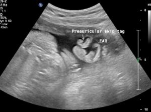
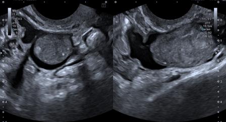
Figure 1: The far lateral parasagittal view of the fetal head revealed unilateral preauricular lobulated soft tissue structures, suggestive of skin tags.

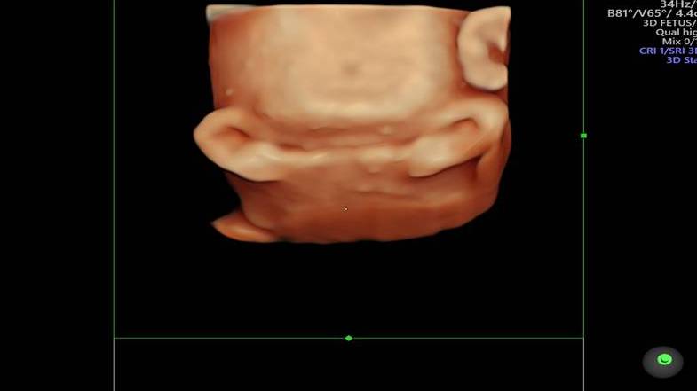
Figure 2: The 3D-Ultrasound image clearly depicts the preauricular skin tags.

Figure 3: The 3D-Ultrasound image clearly depicts the preauricular skin tags.
Careful examination did not reveal any other structural anomaly. Hence, a diagnosis of isolated unilateral preauricular skin tags was made.
Post-natal follow-up confirmed the finding of unilateral preauricular skin tags. The baby was otherwise normal.

Figure 4: Post-natal picture shows the unilateral preauricular skin tags.
Preauricular skin tags are congenital anomalies which consist of lobulated or pedunculated benign skin masses located anterior to external ear. They can be unilateral or bilateral and single or multiple.
Imperfect fusion of 1st and 2nd branchial arches is the underlying etiology of this anomaly. It is relatively common and the incidence is 5 to 10 per 1000 live births.[1] Preauricular skin tags can be an isolated finding or maybe associated with other conditions. When isolated, they don’t have much significance and are of only cosmetic concern.
Risk of chromosomal anomalies is relatively high, mainly Trisomy 13 and, to a lesser extent, Trisomy 18. A very high association has been found with non-chromosomal syndromes and other external ear anomalies. Some associated syndromes are Goldenhar syndrome, Cat eye syndrome, Fraser syndrome, Nager syndrome and Treacher Collins syndrome.[2] Association with renal anomalies like renal agenesis, cystic kidneys, hydronephrosis, duplex collecting system, vesico-ureteric reflux etc. has been reported, but studies show conflicting results.[2]
The diagnostic view on ultrasound is the most lateral parasagittal view of fetal head. Another useful view is the axial view at the level of the orbit.[2] 3D USG is an extremely useful tool for demonstration of external ear anomalies.
Once an external ear anomaly is detected in the fetus, a careful evaluation of facial anatomy is necessary to rule out any facial dysmorphism. Additionally, a detailed anomaly scan needs to be performed to rule out any other associated anomalies.[2]
© Copyright Reserved
References:
1. Dancel R, Price D, Kaufmann L. Evaluation of newborns with preauricular skin lesions. Am Fam Physician. 2012 May 15;85(10):993-8. PMID: 22612052.
2. Paladini D, Volpe P. Ultrasound of congenital fetal anomalies: differential diagnosis and prognostic indicators. 2nd Ed. Boca Raton, Florida: Taylor & Francis Group; 2014.