A very rare antenatal ultrasound diagnosis: cyclopia-otocephaly-agnathia-synotia-astomia complex

A 25-years old primigravida presented for obstetric ultrasound at 19 weeks of gestation. She had a non-consanguineous marriage, no history of abortion, diabetes, hypertension or any drug intake.
The ultrasound findings were as follows –
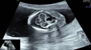
Figure 1: Small fetal head with monoventricle.
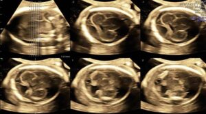
Figure 2: Tomographic ultrasound imaging shows absence of midline falx and fusion of thalami diagnostic of alobar holoprosencephaly.
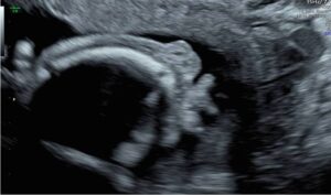
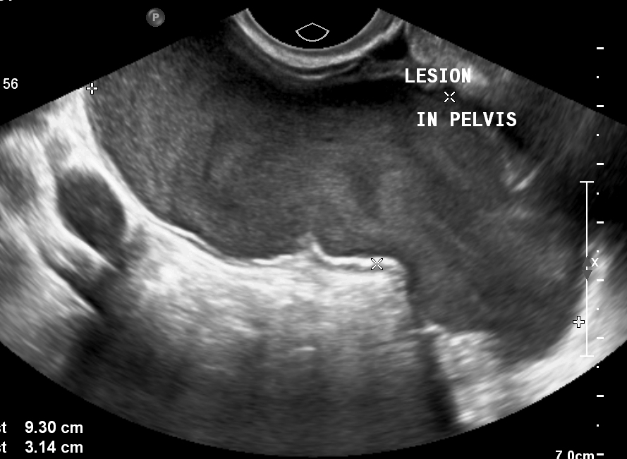
Figure 3: Transvaginal ultrasound revealed orbits fused in the midline (cyclopia).
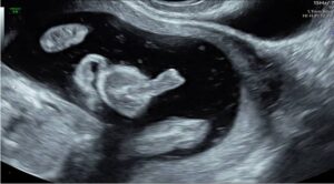
Figure 4: A midline soft-tissue protuberance (proboscis) seen at the level of mid-face. No evidence of mouth, mandible & nose.
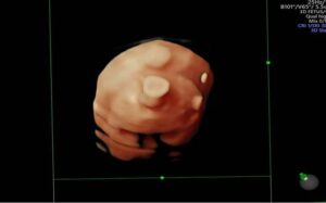
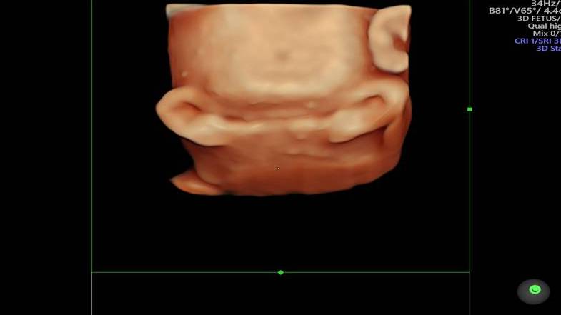
Figure 5: 3D rendering shows cyclopia & proboscis. Absence of mouth and mandible.

Figure 6: 3D rendering demonstrates both ears fused in the midline just below the face (synotia).
Based on the ultrasound findings a diagnosis of cyclopia-otocephaly-agnathia-synotia-astomia complex was made.
The pregnancy was terminated. Following appearance was seen after termination –
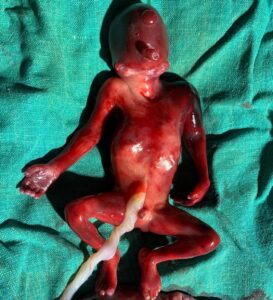
Figure 7: The terminated fetus reveals cyclopia-otocephaly-agnathia-synotia-astomia complex.
Among the complex of anomalies in this case, the most striking and exceedingly rare finding is otocephaly.
Otocephaly is a lethal congenital disorder that occurs due to failure of development of first branchial arch. It is characterised by absence of mandible with fusion of ears in the midline below the chin (synotia). Commonly associated anomalies include astomia / microstomia, aglossia / microglossia, cyclops with proboscis, median cleft lip and cleft palate.[1] The commonest associated neurological anomaly is holoprosencephaly.[1] Other associations include situs inversus, cardiac and genitourinary anomalies.[1]
Failure of the separation of embryonic prosencephalon into two cerebral hemispheres results in holoprosencephaly. It is of three types – lobar, semilobar and alobar. The alobar variety is the most severe, characterised by single midline monoventricle, fused thalami, absent midline structures (interhemispheric fissure, falx, septum pellucidum, third ventricle) and agenesis or hypoplasia of corpus callosum. DeMyer et al described five types of fetal facies in holoprosencephaly. Type 1 consists of cyclopia, in which case a single eye is located in the place normally occupied by root of nose, associated with arhinia (absent nose) and a proboscis.[2]
Cyclopia is the most severe facial presentation of holoprosencephaly. It results in a single midline orbit that could be monopthalmic, synopthalmic or anopthalmic.[3]
A severe case of otocephaly with cyclopia, agnathia, astomia and synotia was described by Indian neonatologist Kanwar Singh and his associates in 2018. The name Kanwar syndrome was assigned by them for this presentation.[3]
© Copyright Reserved
References:
1. Agarwal S, Sen J, Jain S, Rathi SK. Otocephaly: Prenatal and postnatal imaging findings. J Pediatr Neurosci. 2011 Jan;6(1):94-5. doi: 10.4103/1817-1745.84426. PMID: 21977107; PMCID: PMC3173934.
2. Wai LT, Chandran S. Cyclopia: isolated and with agnathia-otocephaly complex. BMJ Case Rep. 2017 Aug 30;2017:bcr2017220159. doi: 10.1136/bcr-2017-220159. PMID: 28855214; PMCID: PMC5589031.
3. Singh K, Sharma S, Agarwal K, Kalra A. Cyclopia-otocephaly-agnathia-synotia-astomia complex: A case report. J Clin Neonatol 2018;7:177-80.