Is this an IUCD or something else?

A 32-year old female presented with dysmenorrhoea for an ultrasound examination. The lady was also additionally being investigated for secondary infertility. There was a past history of repeated medical termination of pregnancies.
A transvaginal ultrasound examination revealed the following:
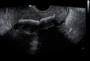
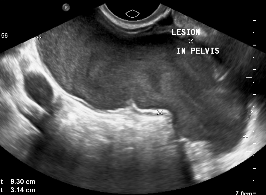
Figure 1: Irregular, linear hyperechoic area with posterior acoustic shadowing seen within the endometrial cavity on sagittal image.
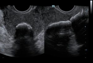
Figure 2: The transverse and sagittal images demonstrate the plate-like hyperechoic content within the endometrial cavity.
Figure 3 – video: The hyperechoic content occupying the endometrial cavity.
Based on the ultrasound findings and clinical presentation, a diagnosis of endometrial osseous metaplasia was made.
Hysteroscopic removal of the lesion was performed and histopathology confirmed mature bony trabeculae with proliferative endometrium in the lesion. At the cellular level, this condition is non-epithelial metaplasia.
The etiopathogenesis is equivocal. Possible mechanisms that have been described include –
(a) Osseous metaplasia of endometrial stromal cells due to chronic endometrial inflammation; (b) Dystrophic ossification of the products of conception after an abortion; (c) Retained fetal tissue promoting osteogenesis in the endometrium; (d) Chronic endometrial inflammatory conditions like pyometra etc; (e) Metabolic disorders. [1]
There is a strong association between this condition and the history of abortions. It may take anywhere between eight weeks to several years after the abortion for the bone formation to manifest. [2]
Infertility associated with endometrial osseous metaplasia is due to its action similar to intra-uterine contraceptive device (IUCD). [3] In fact, IUCD is the main sonographic differential diagnosis of this condition. In the Indian setting, calcification associated with endometrial tuberculosis should also be considered.
© Copyright Reserved
References:
1. Patil SB, Narchal S, Paricharak DG, More SS. Endometrial Osseous Metaplasia: Case Report with literature review. Ann Med Health Sci Res 2013;3 (Suppl1):S10-S12.
2. Cayuela E, Perez-Medina T, Vilanova J, Alejo M, Canadas P. True osseous metaplasia of the endometrium: the bone is not from a fetus. (e1-4).Fertil Steril. 2009;91(1293):1
3. Garg D, Bekker G, Akselroad F, Narasimhulu DM. Endometrial osseous metaplasia: an unusual cause of infertility. BMJ Case Rep 2015; 2015: bcr2015209523.